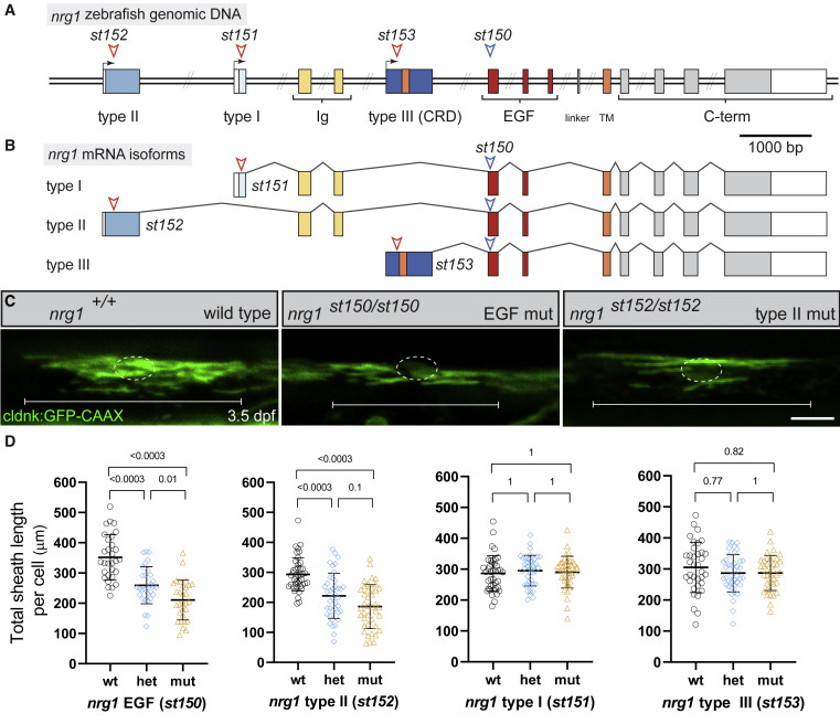
Fig. 1 nrg1 type II signals are required for normal myelination in the spinal cord
(A) Diagram of the zebrafish nrg1 locus. Colored rectangles indicate coding exons; open rectangles indicate UTRs. Arrows indicate alternative transcriptional start sites. Exon length is to scale; double slashes indicate intronic regions that are not to scale. Red arrowheads indicate sgRNA targets in 5′ isoform-specific exons and locations of corresponding mutations. Blue arrowhead indicates sgRNA target used to generate st150 mutation in the EGF domain, which is predicted to eliminate function of all nrg1 isoforms.
(B) Diagram of the mRNA splicing of nrg1 isoforms, illustrating the isoform-specific nature of 5′ exons and chosen sgRNAs.
(C) Confocal images of oligodendrocytes in the dorsal spinal cord at 3.5 dpf labeled using the claudink:GFP transgene. Wild-type fish have normal sheath parameters, while oligodendrocytes (OL) in EGF and type II mutants have reduced myelin. A dotted circle indicates the OL cell body, while brackets indicate the breadth of a single OL. Scale bar, 20 μm.
(D) Quantification of total myelin sheath length produced by individual oligodendrocytes in nrg1 EGF and isoform-specific lines. OLs in nrg1 EGF and type II mutants (mut) make significantly less myelin per OL than in wild-type (wt) animals, while no difference is observed in nrg1 type I or III mutant animals. Animals were genotyped after imaging. Error bars represent mean ± SD. A t test was used to assess significance, and p values were adjusted for multiple comparisons. Each point represents one OL; ≥ 30 OLs per category from ≥4 animals per category; one representative experiment shown from three replicates. Figures 1A and 1B modified from Lysko et al.34 See also Figures S1 and S2.

