
Fig. 9.
- ID
- ZDB-IMAGE-230420-149
- Publication
- Kugler et al., 2023 - GliaMorph: A modular image analysis toolkit to quantify Müller glial cell morphology
- All Figures
- Figures for Kugler et al., 2023
|
Fig. 9.
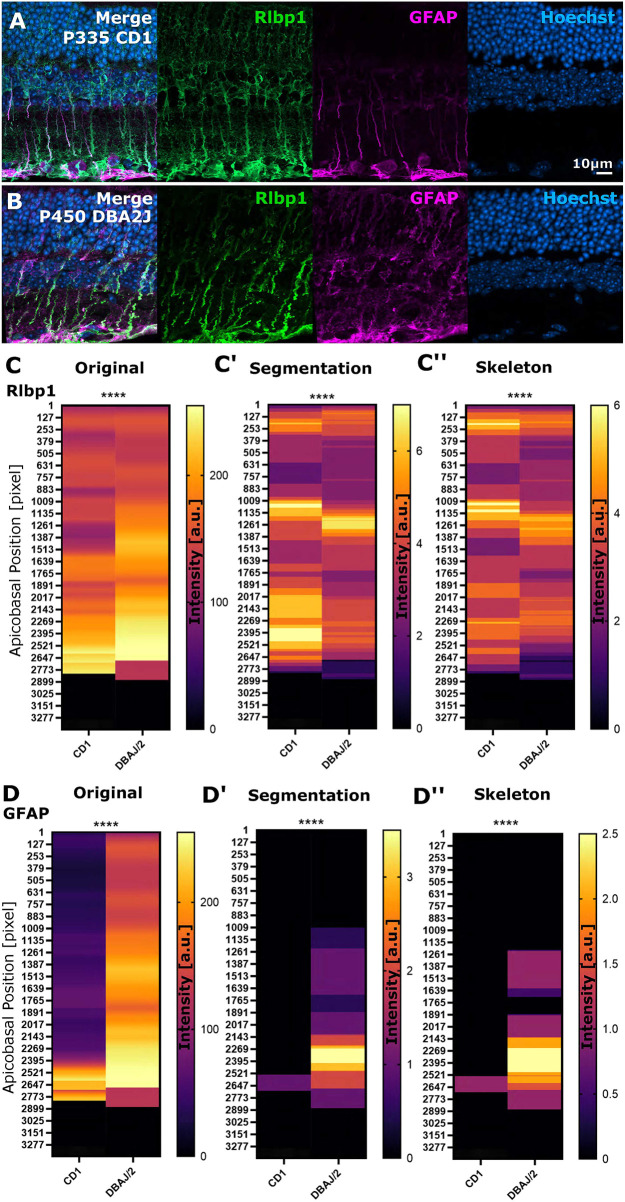
Apicobasal texture analysis identifies subcellular changes in protein localization in mouse glaucoma models. (A,B) Micrographs of stainings in controls and spontaneously glaucomatous DBA/2/J mice (Rlbp1 labels MG, GFAP labels MG reactivity, DAPI labels cell nuclei). (C-C″) Apicobasal texture analysis of Rlbp1 using the zonationTool in original, segmented and skeletonized data showed significant structural changes in glaucoma (original ****P<0.0001; segmentation ****P<0.0001; skeleton ****P<0.0001). (D-D″) Apicobasal texture analysis of GFAP using the zonationTool in original, segmented and skeletonized data showed significant subcellular expression changes in glaucoma models (original ****P<0.0001; segmentation ****P<0.0001; skeleton ****P<0.0001). Mann–Whitney U-test; n=7 stacks from 3 mice each. a.u., arbitrary unit.