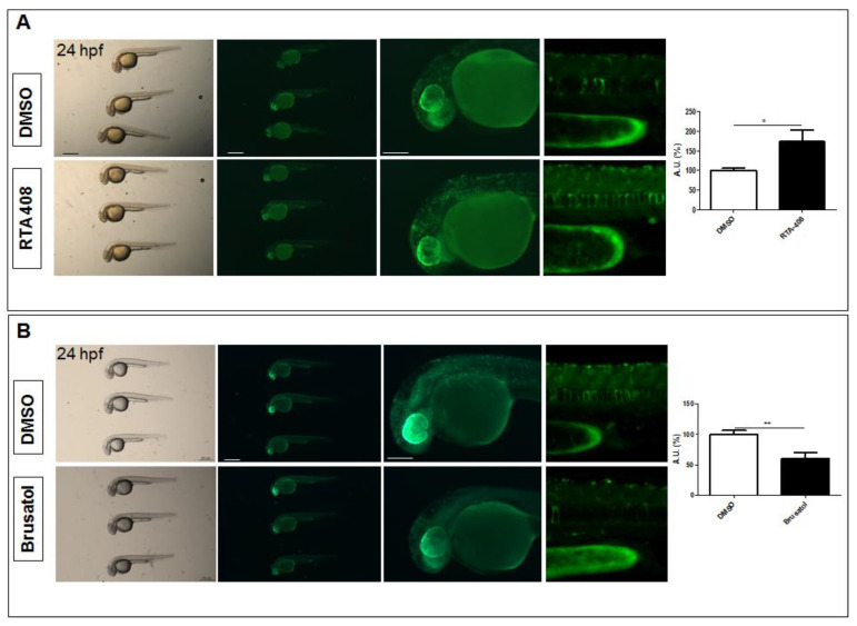
Figure 2
Pharmacological validation of Nrf2/ARE reporter fish. (A) Whole-mount bright field and fluorescence microscopy acquisition of a 24 hpf transgenic larva treated with DMSO and the Nrf2 pathway agonist, RTA-408, for 16 h. A significant increase in fluorescent cells is detected in RTA-408-treated larvae when compared to DMSO-treated fish. (B) Whole-mount bright field and fluorescence microscopy acquisition of a 24 hpf transgenic larva treated with DMSO and the Nrf2 pathway antagonist, Brusatol, for 16 h. A visible decrease in reporter fluorescent intensity is visible in Brusatol-treated larvae. All images are lateral views with anterior to the left. The magnifications of the trunk regions are confocal Z-stack acquisitions. The graphs reported on the right depict the ImageJ-based quantification of the selected trunk region of 5 independently treated fish (* p < 0.05; ** p < 0.005, t-test). Scale bars in the panels with multiple fish: 500 μm; scale bars on the panels with a single magnified larva: 100 μm.

