
|
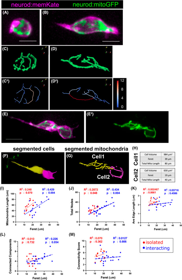
Fig. 4
Mitochondrial morphologies of clustering islet cells. (A, B) Maximum intensity projection of image stack of isolated islet cells from neurod:mitoGFP;neurod:memKate transgenics at 7 dpf. Mitochondrial network (C, D) and skeleton (C′, D′) as analysed by