
|
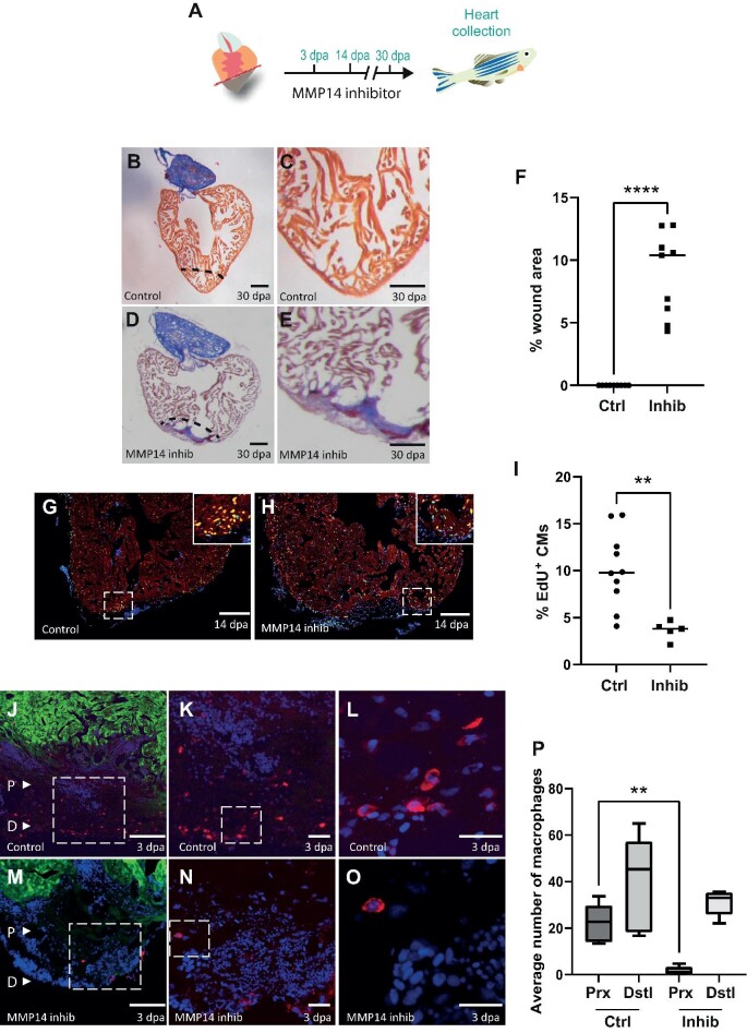
Figure 5
MMP14-expressing macrophages are required for cardiac regeneration. (A) Schematic of the inhibitor treatment protocol. (B and D) AFOG staining of a control (B) or NSC405020-treated (D) heart at 30 dpa. The black dashed line indicates the plane of amputation. Scale bar, 200 μm. (C and E) The same images as in B and D are shown at higher magnification, respectively. Note the presence of a large fibrin (red)/collagen (blue) scar. Scale bar, 100 μm. (F) The graph indicates the average area of the wound region as a percentage of the entire ventricle (n = 9). (G and H) EdU labelling of cycling cardiomyocytes in a control (G) or NSC405020-treated (H) heart. MEF2/cardiomyocytes (red), EdU (green). The dashed white box indicates the area shown at higher magnification in the inset. Scale bar, 200 μm.