Figure 4
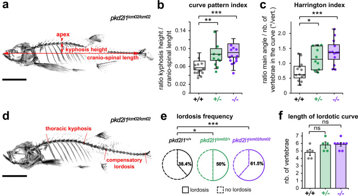
Adult pkd2l1 mutants develop a sagittal imbalance of the spine. (a) Representative 3D micro-computed tomography reconstruction of a pkd2l1icm02/icm02 mutant (18 months-old) used to exemplify the evaluation in the sagittal plane of the kyphosis height, the cranio-spinal length and the kyphosis apex. Scale bar 0.5 cm. (b, c) Distribution of the curve pattern index (b) and Harrington index (c) in wild-type (black, + / + , n = 13), heterozygous (green, + /−, n = 10) and pkd2l1icm02/icm02 mutants (purple, −/−, n = 13). Boxplots represent median values ± IQR. Each point represents a single animal. *: p < 0.05, **: p < 0.01, ***: p < 0.001, Mann–Whitney test. (d) Representative 3D micro-computed tomography reconstruction used to identify in the sagittal plane the thoracic kyphosis and a compensatory lordosis in the caudal region of the spine, exemplified in a 18 months-old pkd2l1icm02/icm02 mutant. Scale bar: 0.5 cm. (e) Pie charts representing the frequency of caudal lordotic curves in wild-type (black, n = 13), heterozygous (green, n = 10) and pkd2l1icm02/icm02 mutants (purple, n = 13). *: p < 0.05, ***: p < 0.001, Chi-Square test. (f) Histogram representing the length of the compensatory curve in lordotic wild-type (grey, + / + , n = 5), heterozygous (green, + /−, n = 5) and pkd2l1icm02/icm02 animals (purple, −/−, n = 6). Each point represents a single animal. Histograms represent mean values ± SEM. ns: p > 0.05, Mann–Whitney test.

