
Figure 3
- ID
- ZDB-IMAGE-230406-19
- Publication
- Marie-Hardy et al., 2023 - Loss of CSF-contacting neuron sensory function is associated with a hyper-kyphosis of the spine reminiscent of Scheuermann's disease
- All Figures
- Figures for Marie-Hardy et al., 2023
|
Figure 3
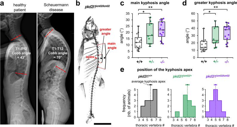
Pkd2l1 loss-of-function induces a hyper-kyphosis of the thoracic spine reminiscent of Scheuermann’s disease. (a) X-ray radiographs in the sagittal plane of a healthy patient (left; male, 20 years old) and of a patient with Scheuermann’s idiopathic disease (right; male, 20 years old). The deformity of the thoracic spine (right) is characterized by a Cobb angle between the first (T1) and the twelfth (T12) thoracic vertebra of 70° and is associated with no vertebral malformations. (b) Representative 3D micro-computed tomography reconstructions of a pkd2l1icm02/icm02 mutant (18 months-old) used to exemplify the evaluation in the sagittal plane of the main kyphosis angle, the greater kyphosis angle and the kyphosis apex. Scale bar: 0.5 cm. (c, d) Distribution of the main kyphosis angle (c) and greater kyphosis angle (d) in wild-type (black, + / + , n = 13), heterozygous (green, + /−, n = 10) and pkd2l1icm02/icm02 mutants (purple, −/−, n = 13). Boxplots represent median values ± IQR. Each point represents a single animal. *: p < 0.05, **: p < 0.01, Mann–Whitney test. (e) Distribution of the position of the apex of the kyphotic curve. The average position of the kyphosis apex is represented for each genotype (arrow, mean ± SEM).