
IMAGE
Fig. 9
- ID
- ZDB-IMAGE-230405-15
- Genes
- Publication
- Tan et al., 2022 - Pax2a, Sp5a and Sp5l act downstream of Fgf and Wnt to coordinate sensory-neural patterning in the inner ear
- All Figures
- Figures for Tan et al., 2022
Image
|
Figure Caption
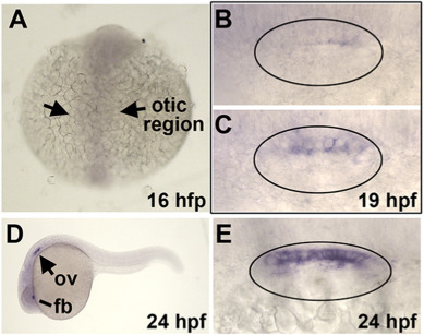
Fig. 9 Expression of sp5b. (A) Dorsal view showing absence of sp5b staining at 16 hpf. The otic region is indicated. (B, C) Dorsal views (anterior to the left) showing variable weak expression of sp5b in the medial wall of the otic vesicle at 19 hpf. (D) Lateral view showing expression of sp5b in the otic vesicle (ov) and forebrain (fb) at 24 hpf. (E) Dorsal view showing sp5b expression in the medial wall of the otic vesicle at 24 hpf.
Figure Data
Acknowledgments
This image is the copyrighted work of the attributed author or publisher, and
ZFIN has permission only to display this image to its users.
Additional permissions should be obtained from the applicable author or publisher of the image.
Reprinted from Developmental Biology, 492, Tan, A.L., Mohanty, S., Guo, J., Lekven, A.C., Riley, B.B., Pax2a, Sp5a and Sp5l act downstream of Fgf and Wnt to coordinate sensory-neural patterning in the inner ear, 139-153, Copyright (2022) with permission from Elsevier. Full text @ Dev. Biol.