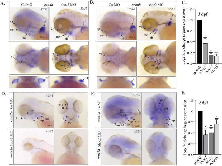
Fig. 6 Extracellular matrix and bone pre-cursor genes are deficient in craniofacial cartilage and bone structures in shoc2 morphants.
Lateral and ventral views of control and shoc2 morphant embryos show expression of the extracellular matrix proteoglycan acana (A) and acanb (B) at 3 dpf. Arrows indicate sites of reduced or lost expression. The total number of embryos used in the statistical analysis is indicated on each image. (C) Total RNA was extracted from control and Shoc2 morphant larvae at 3 dpf and levels of shoc2, acana and acanb mRNA expression were quantified by qPCR. gapdh is a control mRNA. The data are presented as the Log2fold change of the mRNA levels in morphant larvae normalized to control. The results represent an average of three biological replicas. Error bars indicate means with SEM. ∗p<0.05, ∗∗p<0.01, ∗∗∗p<0.001 (Student’s t-test). Lateral and ventral views of control and shoc2 morphant embryos show expression of the runx2a (D) and runx2b (E) at 3 dpf Arrows indicate sites of reduced or lost expression. The total number of embryos used in the statistical analysis is indicated on each image. (F) Total RNA was extracted from control and shoc2 morphant larvae at 3 dpf and levels of shoc2, runx2a and runx2b mRNA expression were quantified by qPCR. gapdh is a control mRNA. The results represent an average of three biological replicas. The data are presented as Log2fold change of the mRNA levels in morphant larvae normalized to control. Error bars indicate means with SEM. ∗p<0.05, ∗∗p<0.01, ∗∗∗p<0.001 (Student’s t-test). ep: ethmoid plate. mc: Meckel’s cartilage. cbs: ceratobranchials. pf: pectoral fin. t: trabeculae. d: dentary. ps: parasphenoid. br: branchiostegal ray. op: opercle. mx: maxilla. pq: palatoquadreate. cl: cleithrum. ch: ceratohyal.
Reprinted from Developmental Biology, 492, Norcross, R.G., Abdelmoti, L., Rouchka, E.C., Andreeva, K., Tussey, O., Landestoy, D., Galperin, E., Shoc2 controls ERK1/2-driven neural crest development by balancing components of the extracellular matrix, 156-171, Copyright (2022) with permission from Elsevier. Full text @ Dev. Biol.

