
|
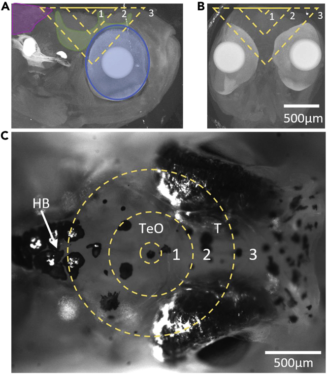
Fig. 6
Illustration of head structures that fall within the cone of light for deep imaging in the adult D. dracula brain
The dashed lines represent the cone of light for an NA of 1 at depths of 100, 400, and 800 μm labeled as 1, 2, and 3, respectively.
(A) Maximum projection of computed tomography (CT) images of the head that contain the right half of the brain. Muscle (magenta), bone (green), and eye pigments (blue) are outlined in the vicinity of the cone of light.
(B) Maximum projection of CT images of the head that contain the brain.
(C) White light image of the adult head. Abbreviations: T, telencephalon; TeO, optic tectum; HB, hindbrain.