
Image
|
Figure Caption
Fig. 6
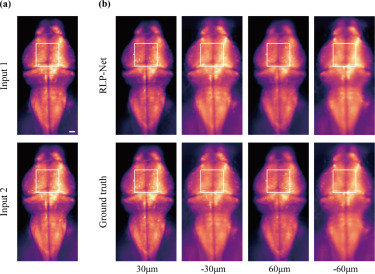
Fig. 6. Virtual refocusing of the images of a larval zebrafish brain. (a) Two adjacent images used as the input to RLP-Net. (b) Refocusing results obtained using RLP-Net (top) and the corresponding ground truth (bottom) at multiple axial locations: = 30, −30, 60, −60 m. The cell bodies of different sets of neurons are in focus at different axial locations in the boxed area. Scale bar, 40 m.
Acknowledgments
This image is the copyrighted work of the attributed author or publisher, and
ZFIN has permission only to display this image to its users.
Additional permissions should be obtained from the applicable author or publisher of the image.
Reprinted from Medical image analysis, 82, Shin, C., Ryu, H., Cho, E.S., Han, S., Lee, K.H., Kim, C.H., Yoon, Y.G., Three-dimensional fluorescence microscopy through virtual refocusing using a recursive light propagation network, 102600, Copyright (2022) with permission from Elsevier. Full text @ Med. Image Anal.