
Image
|
Figure Caption
Fig. 4
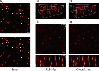
Fig. 4. Virtual refocusing of fluorescent beads. (a) Two adjacent images applied as the input. (b) Oblique maximum intensity projection (MIP) of the reconstructed volume obtained with RLP-Net. (c) As in b, but for the ground truth image. (d) Lateral MIP (top) and axial MIP (bottom) of reconstructed volume. (e) As in d, but for the ground truth image. Scale bars, 50 m.
Acknowledgments
This image is the copyrighted work of the attributed author or publisher, and
ZFIN has permission only to display this image to its users.
Additional permissions should be obtained from the applicable author or publisher of the image.
Reprinted from Medical image analysis, 82, Shin, C., Ryu, H., Cho, E.S., Han, S., Lee, K.H., Kim, C.H., Yoon, Y.G., Three-dimensional fluorescence microscopy through virtual refocusing using a recursive light propagation network, 102600, Copyright (2022) with permission from Elsevier. Full text @ Med. Image Anal.