
Image
|
Figure Caption
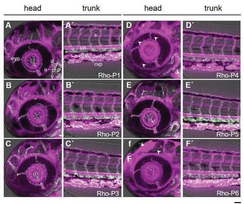
Fig. 5
Biodistribution of the rhodamine labeled peptides (Rho-P1 to Rho-P6) in zebrafish embryos. A,A´) Rho-P1, B,B´) Rho-P2, C,C´)Rho-P3, D,D´) Rho-P4, E,E´) Rho-P5, and f,F,F´) Rho-P6 were intravenously injected into 3 dpf Tg(kdrl:EGFP)s843Tg zebrafish embryos and 3D images from the head (A–F) and trunk (A´–F´) regions were acquired every hour for 19 h (n = 3 embryos for each peptide). f) A single focal plane in the brain region was selected to highlight the distribution of Rho-P6 along the radial glia fiber. Images shown are from 1 h-post-injection. Peptide distribution and blood vessels are shown in magenta and green, respectively. The fluorescence microscopy images are superimposed on bright-field images (grey) from a selected single plane. Lateral views of the embryos are oriented anterior left with dorsal up. a, anterior; p, posterior; d, dorsal; v, ventral; ca, caudal artery; cvp, caudal vein plexus; s, spinal cord. Scale bar: 50 µm.
Acknowledgments
This image is the copyrighted work of the attributed author or publisher, and
ZFIN has permission only to display this image to its users.
Additional permissions should be obtained from the applicable author or publisher of the image.
Full text @ Small