
Image
|
Figure Caption
Fig. 2
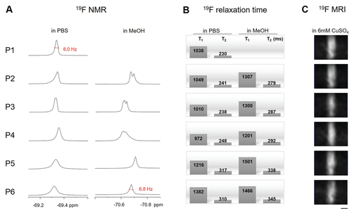
19F characteristics of peptide probes. A) 19F-NMR spectra of peptides P1–P6 at 19F concentration of 5.4 mm (0.2 mm of P1–P4 and 0.6 mm of P5–P6) in PBS buffer at pH 7.4 (left) and methanol (right). The narrowest peak width at half height for each condition is marked in red. B) 19F relaxation times of the peptides (5.4 mm 19F) in PBS and methanol. C) 19F-MRI of peptide solutions in 6 mm CuSO4 (65 mm NaOH added in P1 and P2 solution for pH adjustment) at 19F concentration of 675 mm (P1–P4 at 25 mm and P5–P6 at 75 mm). The aqueous solutions were filled into capillaries with an inner diameter of 1.6 mm and an outer diameter of 2.0 mm, and fitted into a homemade microcoil.[32] Images of coronal planes were acquired using a Fast Low Angle Shot Gradient Echo (FLASH) sequence with a recycle delay of 5.765 ms, an echo delay of 2.72 ms, and a flip angle of 45°. The images were processed using a smoothing algorithm (binning with an average shrink factor 32) of ImageJ. Scale bar: 1 mm.
Acknowledgments
This image is the copyrighted work of the attributed author or publisher, and
ZFIN has permission only to display this image to its users.
Additional permissions should be obtained from the applicable author or publisher of the image.
Full text @ Small