
Image
|
Figure Caption
Figure 1
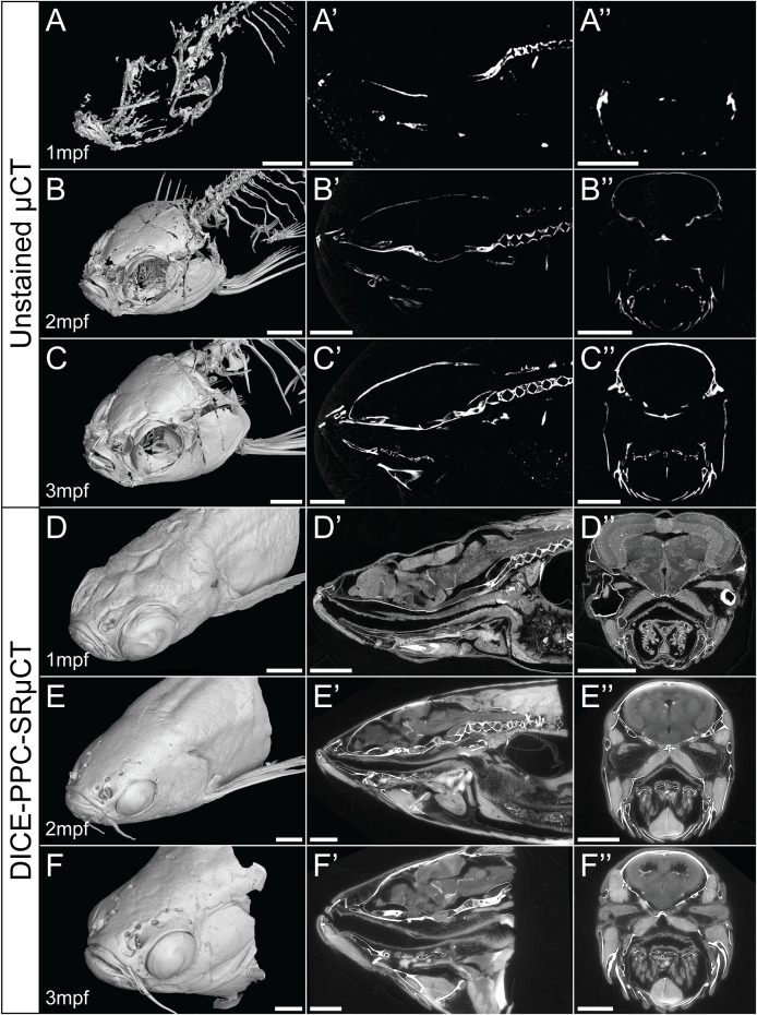
Conventional µCT only visualizes the zebrafish mineralized skeleton, while DICE-PPC-SRµCT adds soft-tissue information. (A-C) Volume renderings of 1-3mpf zebrafish scanned at a voxel size of 5.43µm using conventional µCT. (D-F) Volume renderings of 1-3mpf zebrafish stained with iodine and scanned at 0.727µm voxel size (D) and 3µm (E, F) using PPC-SRµCT. (A’-F’) Sagittal virtual thin sections through the midline. (A’’-F’’) Transverse virtual thin sections through the head (immediately posterior to the eyes). Scale bars, 500µm (A-A’’, D-D’’) and 1mm (B-B’’, C-C’’, E-E’’, F-F’’).
Acknowledgments
This image is the copyrighted work of the attributed author or publisher, and
ZFIN has permission only to display this image to its users.
Additional permissions should be obtained from the applicable author or publisher of the image.
Full text @ Front Endocrinol (Lausanne)