
|
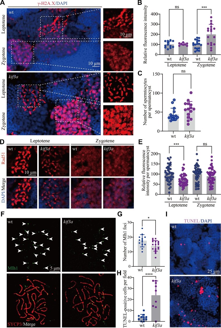
Fig. 4
The repair of DSBs is compromised in the absence of spermatocyte cilia. (A) Confocal images showing the staining of γ-H2A.X (red) in the testis of wild type and Tg(kop:cas9-UTRnanos3;U6:3×sgRNA-kif3a) double transgenic fish. (B) Statistical analysis showing relative fluorescence intensity of γ-H2A.X staining on leptotene and zygotene spermatocytes of wild type and double transgenic fish. (C) Statistical analysis of the number of spermatocytes per spermatocyst in wild type and double transgenic fish. (D) Staining of Rad51 in the testis of wild type and double transgenic fish. (E) Statistical analysis of relative fluorescence intensity of Rad51 staining on leptotene and zygotene spermatocytes. (F) Staining of Mlh1 in primary spermatocytes of wild type and double transgenic fish. (G) Statistical analysis of the Mlh1 foci number per spermatocyte. (H and I) Apoptotic cells stained by TUNEL assay in wild type and double transgenic fish. *P < 0.05; ***P < 0.001; ****P < 0.0001. ns, not significant; wt, wild type.