
|
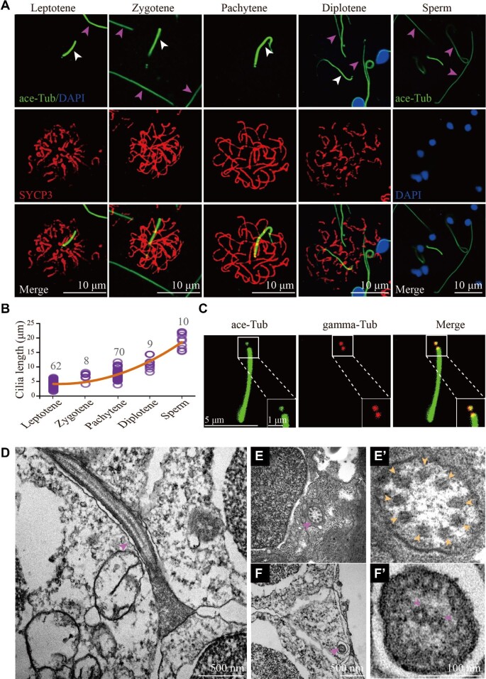
Fig. 1
Cilia of primary spermatocytes in meiosis. (A) Confocal images showing cilia (white arrowhead) and sperm flagella (pink arrowhead) labelled with anti-acetylated tubulin (ace-Tub, green) antibody. The synaptonemal complexes were labelled with SYCP3 (red) and nuclei were counter-stained with DAPI in blue. (B) Statistical results showing the length of cilia in different stages of primary spermatocytes and sperms. (C) Confocal images showing cilia and basal bodies labelled with anti-acetylated tubulin (ace-Tub, green) and anti-gamma tubulin (gamma-Tub, red) antibodies on primary spermatocyte. (D–F’) Transmission electron microscopy results showing the ultrastructure of primary spermatocyte cilia (D–E’) and sperm flagella (F and F’). Cross section showing the ‘9 + 0’ configuration of spermatocyte cilia (E’).