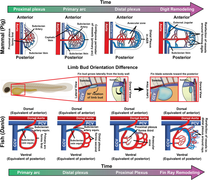
Fig. 9
Schematic of pectoral fin vascular development. Schematics comparing broad stages of forelimb vasculature development in zebrafish and mammals. (A) In mammalian forelimbs, vascular development begins with initial growth of a limb bud-associated capillary plexus and formation of a single predominant arterial feed, the subclavian artery, followed by assembly of a complete circumferential vessel draining to the subclavian vein (and eventually into the cardinal vein). This primitive network expands as the limb grows out, elaborating mid-limb and distal (AER-associated) vascular plexuses separated by an avascular zone. Finally, the distal-most portion of the limb vasculature remodels around the forming digits. (B) In comparison to mammals, the fish limb bud is rotated 90° so that the anterior portion of the fin bud is pointed dorsally instead (the z-axis, from a lateral view of the embryo as in B). A, anterior; P, posterior. (C) In the zebrafish pectoral fin, a circumferential vessel forms first, drawing arterial blood from the dorsal aorta as in mammals but draining into the CCV rather than the (posterior) cardinal vein as in mammals. This circumferential vessel (the primary arc) is analogous to the subclavian artery and vein of mammals. Next, a distal AER-associated plexus forms followed closely by formation of a proximal plexus, leaving a mid-limb avascular zone as in mammals. Lastly, the distal plexus remodels to generate the fin-ray vasculature. PCV, posterior cardinal vein.

