
|
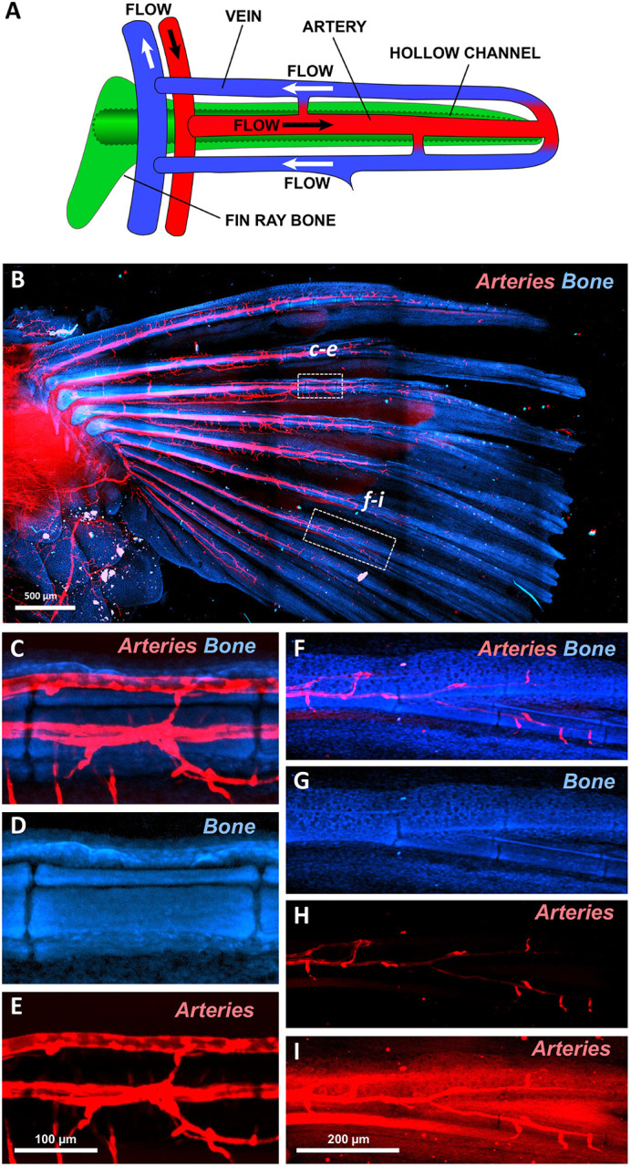
Fig. 2
Fin ray-associated vasculature of the adult zebrafish pectoral fin. (A) Schematic illustrating circulatory flow through an individual adult zebrafish fin ray. Arteries are in red, veins in blue, and fin-ray bone in green. (B-I) Confocal images of an adult pectoral fin dissected from a Tg(kdrl:mcherry)y206 zebrafish, showing Tg(kdrl:mcherry)y20-positive arterial blood vessels (red) and autofluorescent bone (blue). (B) Low-magnification tiled confocal image of a dissected adult pectoral fin. (C-E) Higher-magnification confocal images of the boxed region (c-e) in B, showing Tg(kdrl:mcherry)y206-positive arteries (C,E) running through the center of autofluorescent bones (C,D) in the proximal region of a fin ray. (F-I) Higher-magnification confocal images of the boxed region (f-i) in B, showing weakly Tg(kdrl:mcherry)y206-positive arteries (F,H,I) running through the center of autofluorescent bones (F,G) in the distal region of a fin ray. I shows an ‘overexposed’ image of the fluorescence in H. Images shown are representative of three separate adult fins, with consistent observation of the overall patterning of major vessels. Scale bars: 500 μm (B); 100 µm (C-E); 200 µm (F-I).