
|
Figure 2
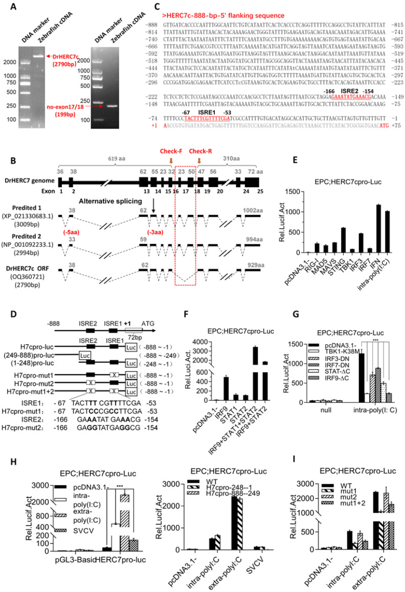
Zebrafish herc7c is a typical ISG. (A) Amplification of full-length cDNA (left) and fragment (right) of the herc7c gene. (B) Exon–intron structure and transcriptional splicing analysis of the zebrafish herc7c genome. The black box represents the exon. The red dotted box indicates the difference area among the possible spliceosomes. The red arrows mark the locations of the check primers. (C) The 5′ flanking sequence of zebrafish herc7c. Two ISRE motifs and the transcription start site are highlighted in red. Their respective positions are numerically marked in the sequence. (D) Schematic diagram of luciferase reporter plasmids under the control of 5′-flanking sequence of zebrafish herc7c gene and the derived truncates and point mutants. (E,F) Zebrafish HERC7c promoter was activated through the RLR signaling pathway and th eJAK-STAT pathway. EPC cells were co-transfected with 200 ng luciferase reporter plasmid (pGL3-Basic or HERC7cpro-Luc), together with 300 ng of each plasmid annotated in the figure. (G) Intracellular poly(I:C) stimulated-activation of zebrafish HERC7c promoter was blocked by overexpression of dominant negative mutants involved in the RLR pathway and the JAK-STAT pathway. (H) Zebrafish HERC7c promoter was activated by intra-poly(I:C), extra-poly(I:C), and SVCV. EPC cells were transfected with 200 ng HERC7cpro-Luc (left panel), or individually with HERC7cpro-Luc and 2 truncated promoter plasmids [(−888~−249) pro-luc, (−248~−1) pro-luc] (right panel). After 12 h, the cells were transfected again with 1 μg/mL poly(I:C) or directly incubated with 50 μg/mL poly(I:C) or infected with SVCV (final titer at 103 TCID50/mL). (I) ISRE motifs were responsible for zebrafish HERC7c promoter activation by intra-poly(I:C). EPC cells were transfected with HERC7cpro-Luc or with 3 ISRE-mutated luciferase plasmids (200 ng each). (*** p < 0.001).