
|
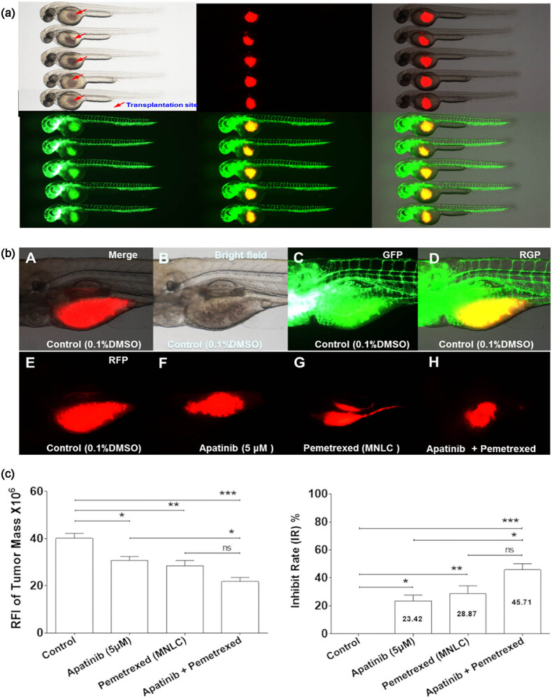
Figure 4
Co-administration of apatinib and pemetrexed inhibited tumor growth in vivo. (a) DiI-stained human A549 cells were successfully grafted into the yolk sac of a zebrafish embryo 2 dpf without immunosuppressant treatment. Approximately 200 cells were injected into the yolk sac and assessed by fluorescence microscopy. (b) Tumor growth at 4 dpi was observed by fluorescence microscopy. (c) Quantitative analysis of inhibitory effects of Apatinib, Pemetrexed, or combination concerning tumor growth. Columns, mean; bars, SEM (n = 10; ANOVA; ***P < 0.0001, **P < 0.01, *P < 0.05, compared to vehicle control group; *P < 0.05, compared to combination group). RFI, relative fluorescence intensity; dpi, days post-injection; MNLC, maximum non-lethal concentration. Zebrafish Strain: fli1a-EGFP; casper. Route of Administration: Soaking in 0.1% DMSO (in fish water).