
|
Figure 3
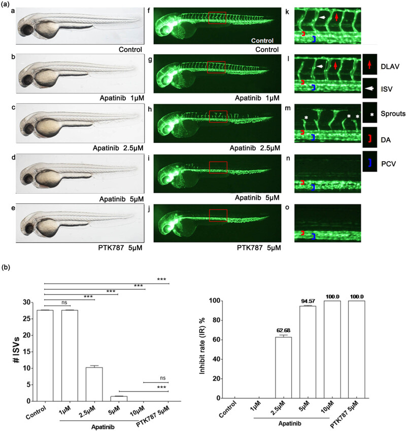
Apatinib inhibits angiogenesis in zebrafish in a dose-dependent manner. (a) (a–o) Representative bright field and fluorescent images of zebrafish embryos at 49 hpf treated with 0.1% DMSO control, apatinib (1, 2.5, 5, and 10 μM), or 5 μM PTK787 (positive control) for 26 h were observed. (f–o) Compared with controls, embryos treated with apatinib presented fewer incomplete ISVs and only occasional sprouts (asterisk) of DA. The boxed regions are shown at higher magnification in the right panels. (b) Quantification of the number of complete ISVs showed a significant decrease in the apatinib-treated embryos. The antiangiogenic effect of apatinib in zebrafish embryos was dose-dependent. # ISVs: The number of complete ISVs (the number of ISVs that connect the DA to the DLAV). Error bars, SEM; ***P < 0.0001 (n = 10; ANOVA); ns, not significant. DLAV, dorsal longitudinal anastomotic vessels; ISV, intersegmental vessels; DA, dorsal aorta; PCV, posterior cardinal vein. Zebrafish strain: fli1a-EGFP; casper. Route of administration: soaking in 0.1% DMSO (in fish water). Animal number: 30 embryos for each condition.