
|
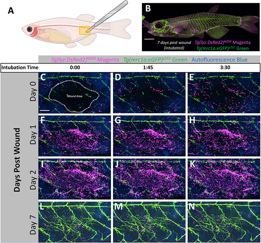
Fig. 4 Repeated intubation and imaging of a small wound. (A) Schematic diagram of an adult casper Tg(lyz:DsRed2)NZ50;Tg(mrc1a:eGFP)y251 double transgenic zebrafish. The approximate site of scale removal by abrasion with a scalpel is noted with a yellow box. (B) An overview image of the intubated fish 7 days post-wounding. Fluorescent neutrophils (magenta), lymphatic vessels (green) and auto-fluorescent scales (blue) are visible. (C-N) Confocal images taken at the beginning (0:00), halfway through (1:45) and at the end of a 3.5 h intubation that was repeated four times (on day 0, 1, 2 and 7). Outline of the wound area is indicated by a dotted line in C. Scale bar: 2.5 mm in B; 500 µm in C. See Movie 2 for full time-lapse sequences.