Fig. 7
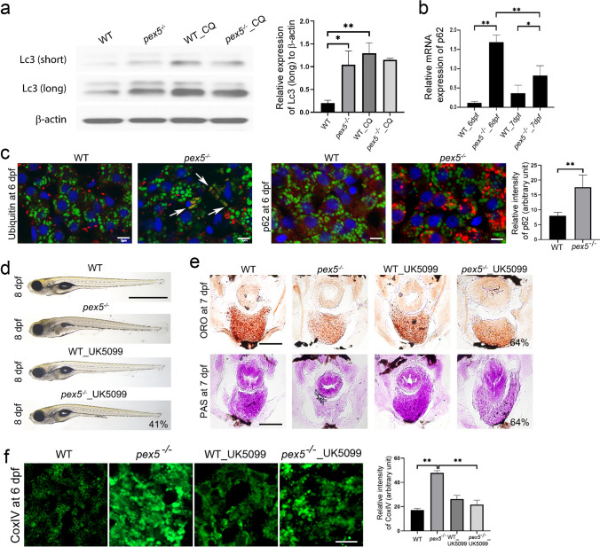
Dysfunctional mitochondria are accumulated in the fasted pex5−/− liver. a Western blotting shows the level of the light chain 3 (Lc3)-II from whole lysates of WT and pex5−/− at 6 dpf with or without chloroquine (CQ, 250 μM). Short and long stand for shorter and longer exposure for film development, respectively. The expression of β-actin was used as the loading control. Graph shows quantified expression levels of Lc3-II (long) relative to β -actin levels and presented as the average of three repeats with error bars indicating standard deviation. Statistical significance was determined using the Student’s t test in Microsoft Excel; * and ** indicate p values < 0.05 and < 0.01, respectively. b Relative expression levels of p62 were determined in reference to those of β-actin by performing quantitative reverse transcription-polymerase chain reaction (qRT-PCR) using total RNA isolated from the liver cells of fasted WT and pex5−/− zebrafish at the indicated stages. Data are presented as the average of three repeats for the relative ratio of p62/b-actin expression. Error bars indicate the standard deviation. Statistical significance was determined using the Student’s t test in Microsoft Excel; * and ** indicate p values < 0.05 and < 0.01, respectively. c Immunofluorescence was performed in liver sections from WT and pex5−/− to detect damaged mitochondria using an anti-Ubiquitin antibody or an autophagy cargo protein using anti-p62 antibody in red as indicated. Mitochondria were visualized through the EGFP signal expressed in the transgenic Tg(Xla.Eef1α1:MLS-EGFP) zebrafish. DAPI stains nuclei in blue in each liver section. Representative images were shown from two independent experiments. Scale bar = 5 μm. Graphs show quantified signal intensity of p62 and presented as the average with error bars indicating standard deviation. Statistical significance was determined using the Student’s t test in Microsoft Excel; ** indicates p value < 0.01. d UK5099 (25 μM) was treated in WT and pex5−/− from 3.5 to 8 dpf, and representative images of the resulting larvae at 8 dpf were laterally presented with anterior to the left. The average percentage representing a near-complete rescue from two independent experiments is indicated. Scale bar equals 1 μm. e Liver sections from the larvae treated with UK5099 from 3.5 to 7 pf were stained either ORO or PAS. Representative images are shown and the percentage of rescue in the levels of liver lipids and glycogen is indicated in the panels of the fasted pex5−/− treated with UK5099. Scale bar = 100 μm. f UK5099 (25 μM) was treated in WT and pex5−/− from 3.5 to 6 dpf, and immunofluorescence was performed in liver sections using anti-CoxIV antibody. Representative images are shown. Scale bar = 5 μm. Graphs show quantified signal intensity of CoxIV at the indicated conditions and presented as the average with error bars indicating standard deviation. Statistical significance was determined using the Student’s t test in Microsoft Excel; ** indicates p value < 0.01

