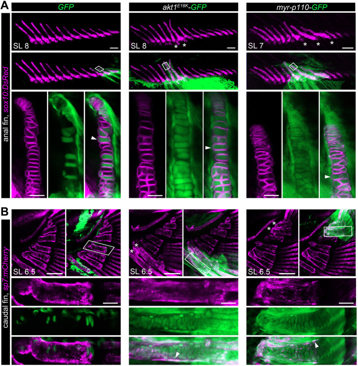
Fig. 2.
Long bone overgrowth is caused by PI3K/AKT overactivation in mesenchymal derivatives. PI3K/AKT mosaic overactivation results in an early overgrowth phenotype in the fin endoskeleton. (A,B) In vivo confocal imaging of chondrocytes (A) marked by Tg(sox10:DsRed) (magenta) and osteoblasts (B) marked by Tg(sp7:mCherry) (magenta) in late larval stages reveals localized skeletal overgrowth in median fins of akt1E18K-GFP (green) and myr-p110-GFP (green) mosaic larva overlapping with GFP+ cells. Boxed areas are at high magnification, showing that akt1E18K-GFP and myr-p110-GFP colocalize with chondrocytes and osteoblasts. Asterisks indicate overgrown bones. Arrowheads indicate GFP+ chondrocytes in A and GFP+ osteoblasts in B. SL, standard length in mm. Scale bars: 100 μm; high-magnification images: 25 μm.

