
|
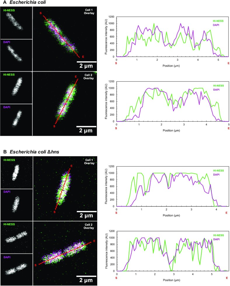
Fig. 1
HI-NESS distribution in wild-type Escherichia coli and E. coli Δhns (Confocal microscopy, single Z-plane). (A) HI-NESS (green) labels the nucleoid in wild-type E. coli where its distribution correlates—albeit poorly—with the DAPI signal (magenta). HI-NESS also distributes in the cytoplasm of these cells, decreasing the signal-to-noise ratio and the applicability of HI-NESS as a DNA label in wild-type E. coli. (B) In E. coli Δhns, HI-NESS localises in the nucleoid. Line scans across E. coli and E. coli Δhns cells are marked in red with start and end positions indicated with S and E, respectively. A white signal in the Overlay images represents colocalization.