Figure 6
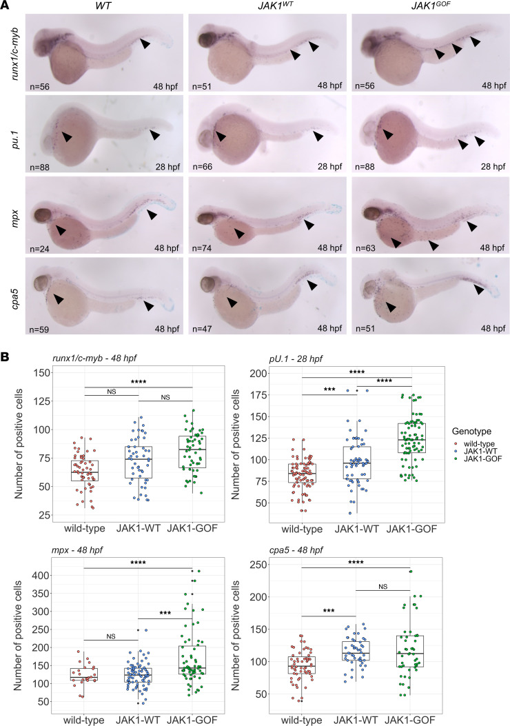
(A) Whole-mount in situ hybridization (WISH) using digoxigenin-labeled RNA antisense probes for stem cells and differentiated myeloid cells in zebrafish embryos of JAK1WT and JAK1GOF transgenic genotypes. A panel of representative micrographs shows runx1/c-myb staining for hematopoietic stem cells at 48hpf, pu.1 staining for early myeloid cells at 28 hpf, mpx staining for neutrophils at 48 hpf, and cpa5 staining for mast cells at 48 hpf. Total numbers of imaged and quantified embryos are shown on the representative images, and arrowheads indicate main sites of marker expression. (B) Plots of marker-positive cell counts for runx1/c-myb at 48 hpf, pu.1 at 28 hpf, and mpx and cpa5 at 48 hpf. Each individual embryo count is indicated by a filled circle, and the box plot shows quartile distribution with whiskers covering 95% CI. Black circles denote location of outlier counts. One-way ANOVA was used to quantify the statistical differences between the groups. ***P ≤ 0.001; ****P ≤ 0.0001.

