Image
Figure Caption
Figure 7
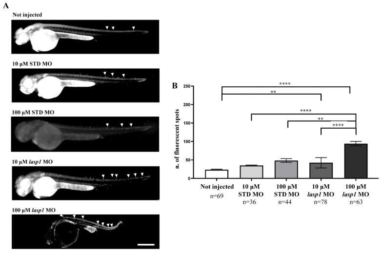
Injection of lasp1 MO increases apoptosis in zebrafish embryos at 48 hpf. (A) Representative images of acridine orange staining by fluorescence microscopy show increased apoptosis in whole embryos following injections of lasp1 MO at 10 µM and 100 µM doses. Fluorescence spots are evidenced by arrowheads. Scale bars: 200 µm. (B) The acridine orange-positive spots were counted using ImageJ. Histograms represent the average numbers of fluorescent spots; error bars are SEMs. ** p < 0.01 and **** p < 0.0001 in one-way ANOVA followed by Bonferroni testing.
Figure Data
Acknowledgments
This image is the copyrighted work of the attributed author or publisher, and
ZFIN has permission only to display this image to its users.
Additional permissions should be obtained from the applicable author or publisher of the image.
Full text @ Genes (Basel)

