
Fig. 3.
- ID
- ZDB-IMAGE-230119-18
- Publication
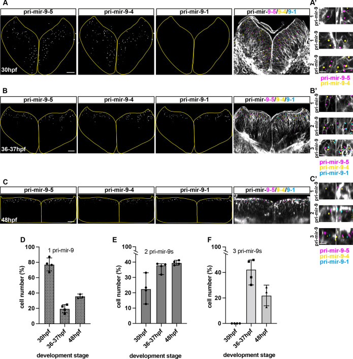
- Soto et al., 2022 - Sequential and additive expression of miR-9 precursors control timing of neurogenesis
- All Figures
- Figures for Soto et al., 2022
|
Fig. 3.