
Image
|
Figure Caption
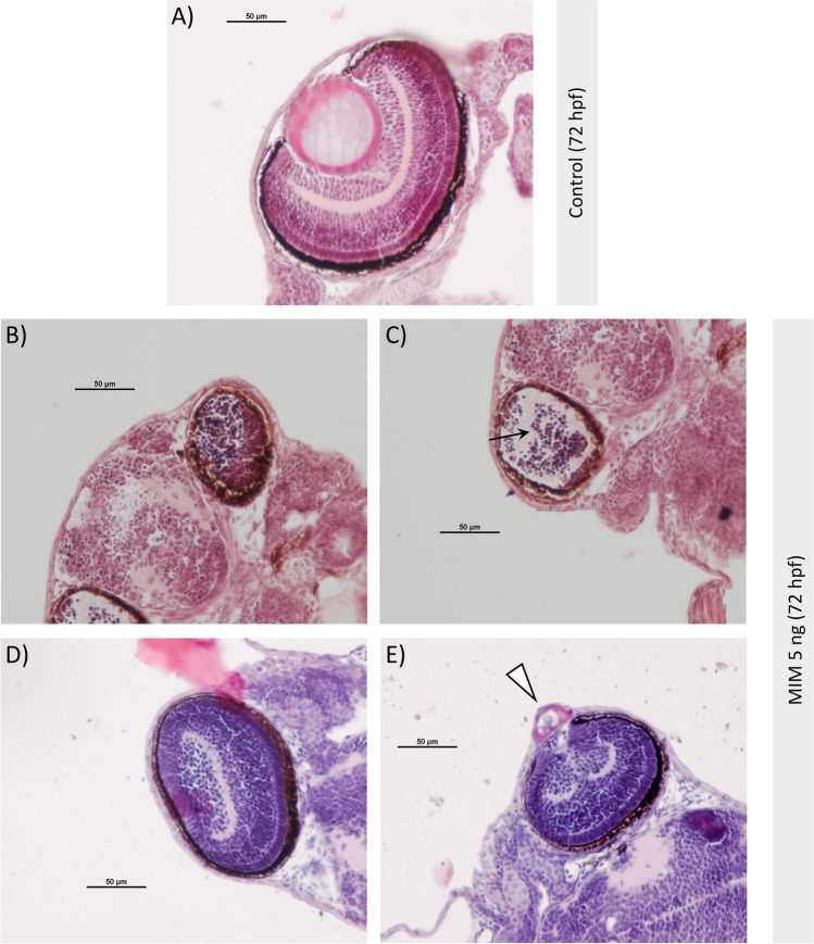
Fig. 4
Morphology of eyes of zebrafish larvae at 72 hpf. A Normal eye structure of larvae from the control group and B–E morphological abnormalities of eyes in larvae microinjected with MiR92b-3p mimic at 5 ng/embryo (MIM 5 ng). The morphological changes in the exposed zebrafish usually manifested as smaller eye diameter, and as a lack of a lens and typical retina stratification (black arrow). In some zebrafish individuals, a vacuolized lens (or its remnants) was located in the delaminated cornea that was expanding outward (white arrowhead)
Acknowledgments
This image is the copyrighted work of the attributed author or publisher, and
ZFIN has permission only to display this image to its users.
Additional permissions should be obtained from the applicable author or publisher of the image.
Full text @ J. Appl. Genet.