
|
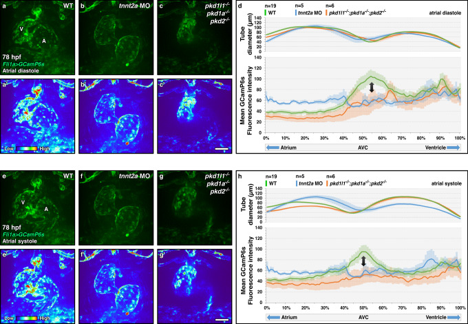
Fig. 4
a–c, e–g Confocal imaging of 78 hpf fli1a:Gal4FF; UAS:GCamP6s wild-type (a, a′, e, e′), tnnt2a morphant (b, b, f, f′) and pkd triple mutant (c, c′, g, g′) hearts during atrial diastole (a–c′) and systole (e–g′); maximum projection of 4D-assembled spinning disc movies. d, h Diameter and endocardial calcium levels along the contracting heart tube in 78 hpf wild-type, tnnt2a morphant, and pkd triple mutant larvae during atrial diastole (d) and systole (h); arrows point to the AV canal calcium peak. Hearts imaged in ventral view, anterior to the top, ventricle (V) on the left, and atrium (A) on the right. Error bars indicate SEM. Scale bars: 50 µm. Source data are provided as a Source Data file.