Fig. 8
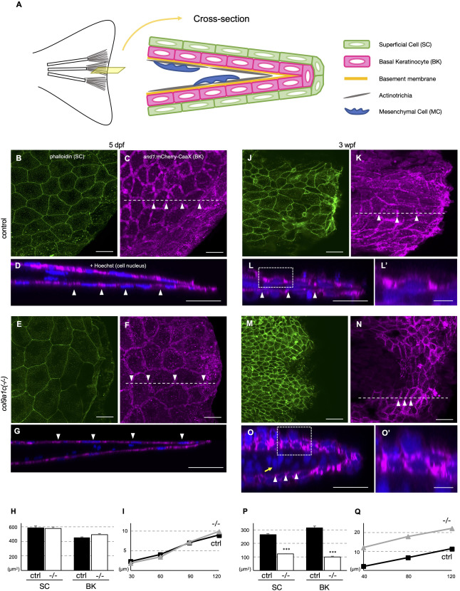
Fig. 8. Orderly arrays of actinotrichia provide a scaffold for epidermal cells to spread. (A) Schematic diagram of the cross-section at the fin tip modified from Kuroda et al. (2018). (B–G) Fin tip region of the control (B to D) and col9a1c(-/-) (E to G) of 5 dpf fish. Cell membrane of superficial cells (SCs) visualized by phalloidin (B and E). Cell membrane of basal keratinocytes (BKs) expressing mCherry-CaaX under the and1 promoter (Kuroda et al., 2018) (C and F). Optical section at the dot line in C and F (D and G). Cell nuclei (blue) were visualized with Hoechst. (H) Cell area of SCs and BKs at 5 dpf (N = 3 each from group). There was no significant difference in either cell. (I) Tissue thickness in D and G. The thickness was measured every 30 μm, starting from the distal tip of the fins. No significant difference in cell thickness between the control and the col9a1c(-/-) mutant. (J–O) Fin tip region of 3-week-old control (J to L) and col9a1c(-/-) fish (M to O). Cell membrane of SCs visualized by phalloidin (J and M). Cell membrane of BKs visualized with mCherry (K and N). Optical sections of the dot line in I and L (L and O). Cell nuclei (blue) were visualized with Hoechst. (L′ and O′) Magnified images of white dashed box in L, O. (P) Cell area of SCs and BKs at 3 wpf (N = 3 each from group). The cell area in col9a1c(-/-) was approximately 50% smaller in SCs and 70% smaller in BKs. (Q) Tissue thickness in L and O. The thickness was measured every 40 μm, starting from the distal tip of the fins. The col9a1c(-/-) mutant showed approximately twice the thickness of tissue compared to the control at every point. White arrowheads in D, G, L, and O indicate the boundary of the two arrayed cells in C, F, K, and N, respectively. Yellow arrow indicates the abnormally oriented nuclei of mesenchyme cells. Scale bars: 20 μm in B–L, M–O; 5 μm in L′, O′.
Reprinted from Developmental Biology, 481, Nakagawa, H., Kuroda, J., Aramaki, T., Kondo, S., Mechanical role of actinotrichia in shaping the caudal fin of zebrafish, 52-63, Copyright (2021) with permission from Elsevier. Full text @ Dev. Biol.

