IMAGE

Fig. 6.

ID
ZDB-IMAGE-221230-34
Source
Figures for Zhang et al., 2023
Image
Figure Caption

Fig. 6.

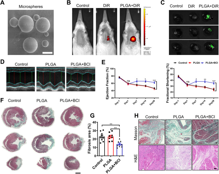
Intramyocardial injection of PLGA-encapsulated BCI improves cardiac function post-MI in rats. (A) Poly (D, L-lactic-co-glycolic acid) (PLGA) microspheres under scanning electron microscopy (scale bar: 5 μm). (B) In vivo fluorescence imaging of rats 4 weeks after intramyocardial injection of normal saline (control), 1,1'-dioctadecyl-3,3,3′,3'-tetramethylindotricarbocyanine iodide (DiR) or PLGA-encapsulated DiR (n=3 per group). (C) Imaging of hearts 4 weeks after intramyocardial injection of normal saline (control), DiR or PLGA-encapsulated DiR (n=3 per group). (D) Representative M-mode tracings from ECHO of control, PLGA-treated and PLGA+BCI treated rat hearts at 4 weeks after MI. (E) Statistical analysis of ejection fraction and fractional shortening of control, PLGA-treated and BCI+PLGA-treated rat hearts 1 day before MI, and on days 1, 7, 14 and 28 after MI (n=8–9). (F) Masson's trichrome staining of control, PLGA-treated and BCI+PLGA-treated hearts at 28 days post-MI. Scale bar: 3 mm. (G) Quantification of Masson's trichrome-stained fibrotic area in F (n=8–9). (H) Masson's trichrome staining and H&E staining of control, PLGA-treated and BCI+PLGA-treated hearts at 28 days post-MI. Scale bars: 300 μm. One-way ANOVA followed by Dunnett's multiple comparison test; mean±s.e.m.; *P<0.05; **P<0.01, ***P<0.001; ns, not significant.

Acknowledgments
This image is the copyrighted work of the attributed author or publisher, and ZFIN has permission only to display this image to its users. Additional permissions should be obtained from the applicable author or publisher of the image. Full text @ Dis. Model. Mech.