
|
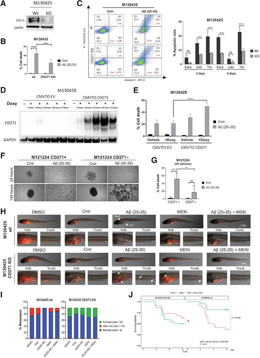
Fig. 5
CD271high cells display a greater susceptibility to Aβ(25–35)-induced death. A, CD271 silencing was confirmed by Western blot. B, Melanoma cells were treated with Aβ(25–35) 40 μmol/L and the amount of dead cells (% sub-G1) was measured by PI staining at the FACS. C, AnnexinV/PI assay was performed to evaluate the percentage of early and late apoptotic cells. D, CMVTO_EV- and CMVTO_CD271-transfected cells were treated with doxycycline, and proteins were collected to evaluate CD271 induction by Western blot. E, Melanoma cells were treated with doxycycline for 48 hours, followed by Aβ(25–35) administration. PI staining was performed 24 hours later and the percentage of dead cells was measured by FACS. F, M121224 CD271+ and − cells were seeded as spheroids and treated with Aβ(25–35). Twenty-four and 144 hours later, brightfield pictures were taken and PI staining (G) was performed. Data represent the mean ± SD of triplicate determinations. H, M130425 wt and CD271 KO cells were injected into the yolk of zebrafish larvae and treated with Aβ(25–35) ± MEKi. Pictures of wt are the same of Fig. 4C. I, The severity of metastasis was evaluated 4 days later by two blind investigators. J, Probability of survival in zebrafish injected with wt versus CD271 KO cells. Two-way ANOVA was used for statistical analysis. **, P < 0.01; ***, P < 0.001; ****, P < 0.00001; ns, nonsignificant.