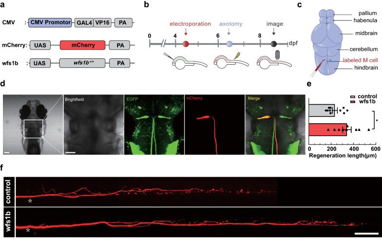
Fig. 5 Complementation of wfs1b at single-cell electroporation promotes M-cell axon regeneration. a Construction of the wfs1b expression system. Plasmids express only mCherry served as the control vector. b Timeline of time points of electroporation, axotomy, and regeneration imaging. c Schematic of M-cell soma electroporation. d Confocal imaging of zebrafish larvae 12 h after electroporation (far left) and magnified images of the brain stem in zebrafish larvae, denoting the position of M-cell soma under different fluorescence in the white box. mCherry represents the axons that were labeled by mCherry. Scale bar, 50 μm. e, f wfs1b gene retro-complementation rescued the length of Mauthner cell axon regeneration (control: 233.1 ± 22.65 μm, n = 10; wfs1b overexpression: 341.4 ± 37.92 μm, n = 9). White asterisk: ablation point. scale bar, 50 μm. P = 0.0225. Assessed by unpaired t test
Image
Figure Caption
Acknowledgments
This image is the copyrighted work of the attributed author or publisher, and
ZFIN has permission only to display this image to its users.
Additional permissions should be obtained from the applicable author or publisher of the image.
Full text @ Acta Neuropathol Commun

