
Figure 7
- ID
- ZDB-IMAGE-221218-20
- Antibodies
- Publication
- Martin et al., 2022 - Proper modulation of AHR signaling is necessary for establishing neural connectivity and oligodendrocyte precursor cell development in the embryonic zebrafish brain
- All Figures
- Figures for Martin et al., 2022
|
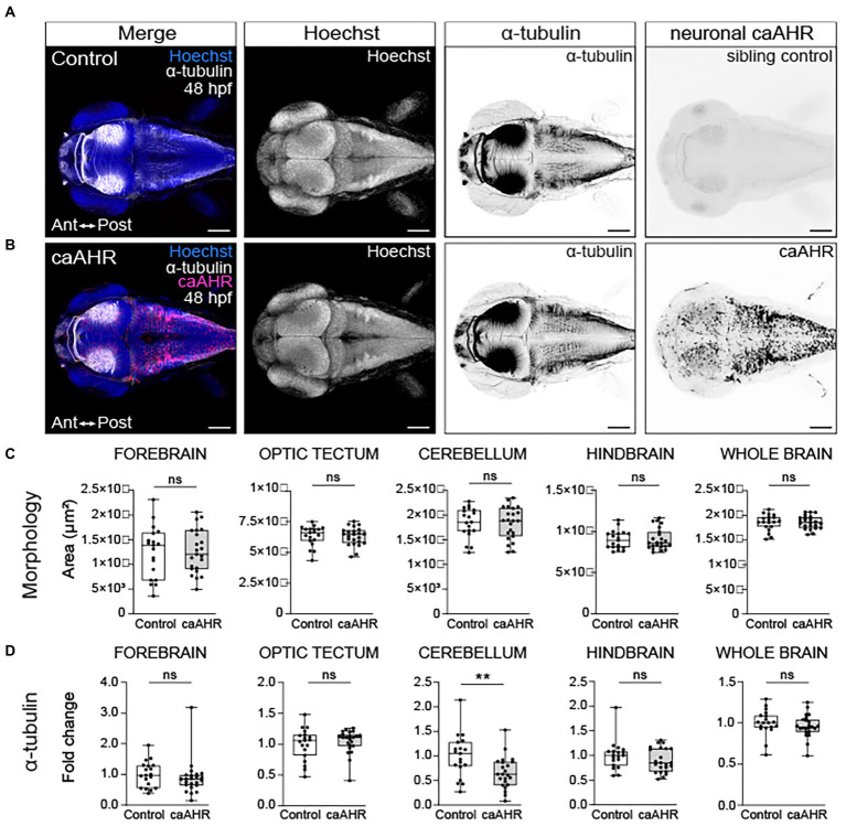
Figure 7
Neuron-specific activation of AHR disrupts the development of the cerebellum without affecting brain regions disrupted by global AHR activation. Confocal images at 48- hpf of control embryos (A) and embryos with neuron-specific expression of a constitutively active AHR [Tg(elav3l:Gal4;UAS:caAHR:2A-tRFP)] (B). Embryos were stained with Hoechst (blue in the first panel and white in second panel of both A,B) to visualize the brain parenchyma and immunolabeled with an anti-acetylated alpha tubulin antibody (α-tubulin) to mark axon tracts (white in the first panel and inverted black in the third panel of both A,B). There were no differences in the morphology of the forebrain, optic tectum, cerebellum, hindbrain, and whole brain at 48 hpf when comparing control (C) embryos to embryos with neuron-specific expression of a constitutively active AHR. (D) Fold change in acetylated α-tubulin expression in the forebrain, optic tectum, cerebellum, hindbrain, and whole brain in control and Tg(elav3l:Gal4;UAS:caAHR:2A-tRFP embryos relative to mean area coverage of α-tubulin in wild-type embryos. Statistical significance determined by Welch’s t-test, *p < 0.05, **p < 0.01, ***p < 0.001. n = 19–23 fish/group across 3 replicates. Anterior (Ant) is left in all confocal micrographs. Scale bar = 100 μm. Magnification = 10x.