
Figure 5
- ID
- ZDB-IMAGE-221218-18
- Antibodies
- Publication
- Martin et al., 2022 - Proper modulation of AHR signaling is necessary for establishing neural connectivity and oligodendrocyte precursor cell development in the embryonic zebrafish brain
- All Figures
- Figures for Martin et al., 2022
|
Figure 5
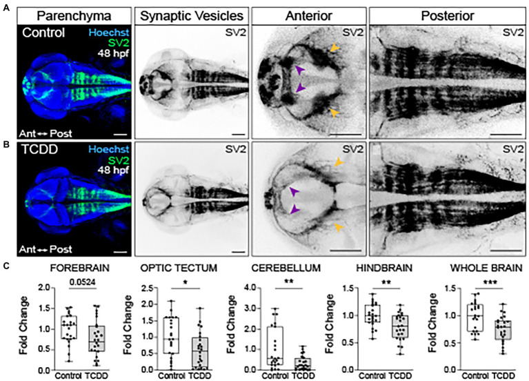
Embryonic TCDD exposure reduces synaptic vesicle 2 immunoreactivity. Dorsal confocal images of control (A) and TCDD-exposed (B) zebrafish at 48 hpf. The brain parenchyma was stained with Hoechst (DNA, blue) and immunolabeled with an antibody marking synaptic vesicle glycoprotein 2 (SV2) (green and inverted black). Forebrain vesicle production was decreased following TCDD exposure with notable reductions in the habenula (purple arrowheads). SV2 immunoreactivity was significantly reduced in the optic tectum (yellow arrowheads), cerebellum and hindbrain, and whole brain following TCDD exposure. (C) Fold change of SV2 immunolabeling in the forebrain, optic tectum, cerebellum, hindbrain, and whole brain was calculated relative to mean SV2 area coverage of controls. Statistical significance was determined by Welch’s t-test, *p < 0.05, **p < 0.01, ***p < 0.001. n = 23–24 fish/group across 3 replicates. Scale bar = 100 μm. Anterior (Ant) is left in all confocal micrographs.