IMAGE

Figure 1

ID
ZDB-IMAGE-221218-14
Publication
Martin et al., 2022 - Proper modulation of AHR signaling is necessary for establishing neural connectivity and oligodendrocyte precursor cell development in the embryonic zebrafish brain
All Figures
Figures for Martin et al., 2022
Image
Figure Caption

Figure 1

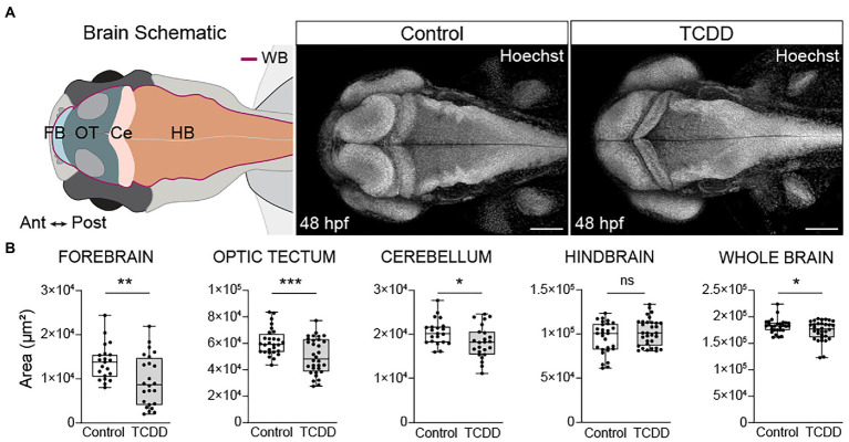
Developmental TCDD exposure impairs embryonic brain morphogenesis. (A) Schematic of the embryonic zebrafish brain at 48 hpf depicting the brain regions quantified in B. Dorsal confocal images of Hoechst (DNA, white) stained embryos and larvae were used to visualize and quantify differences in gross brain morphology. Embryos were exposed at 4 hpf to either control (DMSO) or TCDD (10 ppb) for 1 h. (B) Regional and whole brain size was determined by measuring the area of the forebrain (FB), optic tectum (OT), cerebellum (Ce), hindbrain (HB), and whole brain (WB) in control and TCDD-exposed embryos. Statistical significance determined by Welch’s t-test, *p < 0.05, **p < 0.01, ***p < 0.001. n = 21–26 fish/group across 3 replicates. Anterior (Ant) is left, posterior (Post) is right in all confocal micrographs. Scale bar = 100 μm, magnification = 10x.

Figure Data
Acknowledgments
This image is the copyrighted work of the attributed author or publisher, and ZFIN has permission only to display this image to its users. Additional permissions should be obtained from the applicable author or publisher of the image. Full text @ Front. Mol. Neurosci.