
IMAGE
Figure 5
- ID
- ZDB-IMAGE-221214-57
- Antibodies
- Publication
- Chrispell et al., 2022 - Grk7 but not Grk1 undergoes cAMP-dependent phosphorylation in zebrafish cone photoreceptors and mediates cone photoresponse recovery to elevated cAMP
- All Figures
- Figures for Chrispell et al., 2022
Image
|
Figure Caption
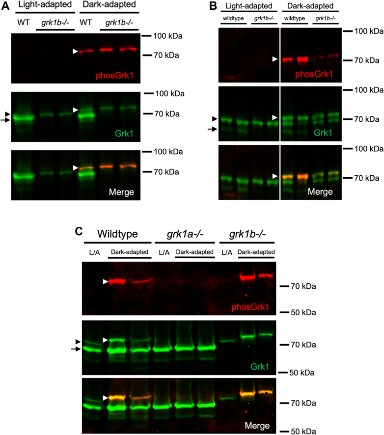
Figure 5
Grk1 phosphorylation in rod grk1a and cone grk1b KO zebrafish. Immunoblots were probed with antibodies against total Grk1 (green) and phosphorylated Grk1 (red), followed by incubation with secondary antibodies. Comparison of Grk1 phosphorylation in (A) light- or dark-adapted WT and grk1b−/− larvae at 5 dpf, (B) light- or dark-adapted WT and grk1b−/− adults, and (C) light- or dark-adapted WT, grk1b−/−, and grk1a−/− larvae at 5 dpf. Black arrowhead: Grk1a; Black arrow: Grk1b; White arrowhead, phosphorylatedGrk1a. Panel B was spliced for clarity due to noncontiguous loading on the same gel.
Figure Data
Acknowledgments
This image is the copyrighted work of the attributed author or publisher, and
ZFIN has permission only to display this image to its users.
Additional permissions should be obtained from the applicable author or publisher of the image.
Full text @ J. Biol. Chem.