
Fig. 1
- ID
- ZDB-IMAGE-221212-31
- Publication
- Bernardello et al., 2021 - Analysis of intracellular protein dynamics in living zebrafish embryos using light-sheet fluorescence single-molecule microscopy
- All Figures
- Figures for Bernardello et al., 2021
|
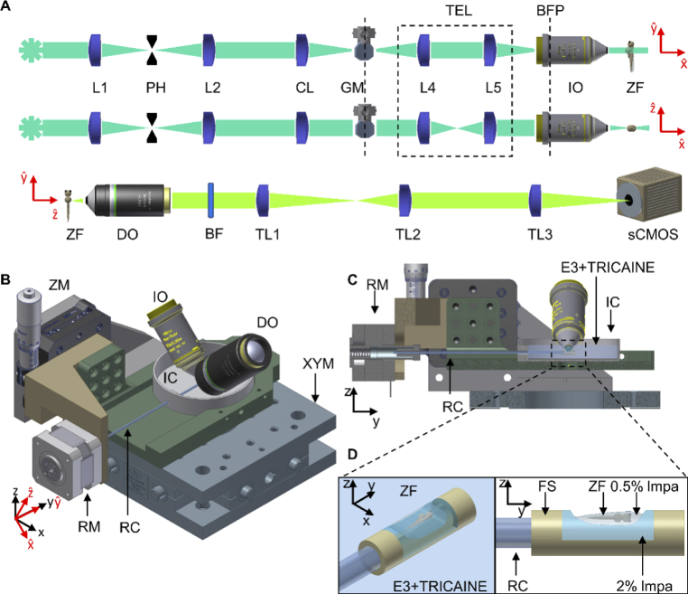
Fig. 1 Schematic overview of the LSFM platform for in vivo SMM imaging. (A) schematic of the optical illumination (top two) and detection (bottom one) light paths; (B) schematic of the sample mounting system, with (C) its lateral section view, (D) the FEP support for zebrafish mounting. L = lens, PH = pinhole, CL = cylindrical lens, GM = galvo-mirror, BFP = back focal plane, TEL = telescope plane-conjugating GM to BFP, IO = Illumination objective, DO = detection objective, BF = bandpass filter, TL = tube lens, ZF = zebrafish, RC = rotation capillary, FS = FEP support, RM = rotational motor, IC = imaging chamber, ZM = motorized stage in z, XYM = motorized stage in xy, lmpa = low melting point agarose.