
Image
|
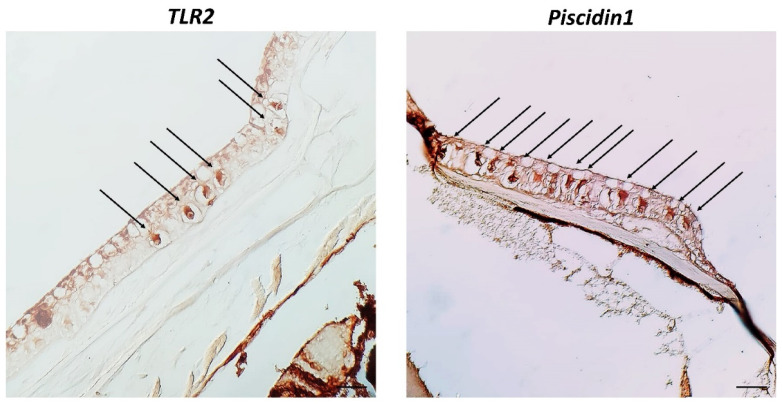
Figure Caption
Figure 3
Cross sections (5 μm thick) of zebrafish skin. Sections are immunohistochemically treated with TLR2 and Piscidin1. Immunoperoxidase 40×, scale bar 40 μm. There are distinct CCs for TLR2 and Piscidin1 (arrows), with a significant core in the middle. They are neatly organized in the middle epidermal layer.
Acknowledgments
This image is the copyrighted work of the attributed author or publisher, and
ZFIN has permission only to display this image to its users.
Additional permissions should be obtained from the applicable author or publisher of the image.
Full text @ Biology (Basel)