
Image
|
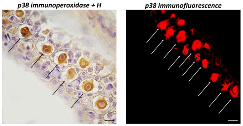
Figure Caption
Figure 2
Longitudinal sections (5 μm thick) of zebrafish skin, Sections are immunohistochemically treated with MAPK p38. Immunoperoxidase counterstained by H, 100×, scale bar 100 μm. Immunofluorescence, 40×, scale bar 40 μm. Immunoreactive CCs for MAPK p38 (arrows) appear evident, with a large core centrally located. They are well organized in the intermediate epidermal layer as shown by counter colouring with H.
Acknowledgments
This image is the copyrighted work of the attributed author or publisher, and
ZFIN has permission only to display this image to its users.
Additional permissions should be obtained from the applicable author or publisher of the image.
Full text @ Biology (Basel)