Image
Figure Caption
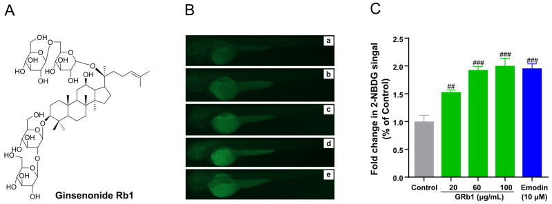
Figure 2 GRb1 induces 2-NBDG uptake in zebrafish larvae. (A) Chemical structures of ginsenoside Rb1. (B) Fluorescence microscopy images of zebrafish larvae treated with different concentrations of GRb1 (20, 60, 100 μg/mL) with 10 μM emodin as a positive control. Control (a), 20 μg/mL GRb1 (b), 60 μg/mL GRb1 (c), 100 μg/mL GRb1 (d) and 10 μM emodin (e). (C) Fold change in 2-NBDG absorption quantified by Image J. Results are expressed as mean ± SEM. ### p < 0.001, ## p < 0.01 vs. control.
Acknowledgments
This image is the copyrighted work of the attributed author or publisher, and
ZFIN has permission only to display this image to its users.
Additional permissions should be obtained from the applicable author or publisher of the image.
Full text @ Metabolites

