
|
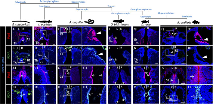
FIGURE 6 Pattern of presence/absence of an asymmetric pax6 dorsal nucleus in actinopterygians. (A-R) Transverse sections of habenulae in the reedfish E. calabaricus [(A,B); 20 cm juvenile], spotted gar L. oculatus [(C,D); 8.5 cm juvenile], European eel A. anguilla [(E–H); yellow resident stage], silver arowana O. bicirrhosum [(I–L); 7 cm juvenile], zebrafish D. rerio [(M,P); adult] and false clownfish A. ocellaris [(Q–T); stage 6], following IHC with antibodies directed against pax6 (A,C,E,I,M,Q,G,K,O,S) and PCNA (B,D,F,J,N,R,H,L,P,T). (A1–H1), (Q1–T1) are higher magnification views of the territories boxed in (A–H), (Q–T). Arrows point towards pax6 and PCNA positive neural progenitors, white arrowheads point to left restricted pax6 positive and PCNA negative dorsal nuclei observed in the European eel and false clownfish as in the Atlantic salmon. Opened arrowheads indicate thalamic Pax6 positive neuronal populations. Scale bars = 100 μm in (A–L) and 50 μm in (M–T).