
|
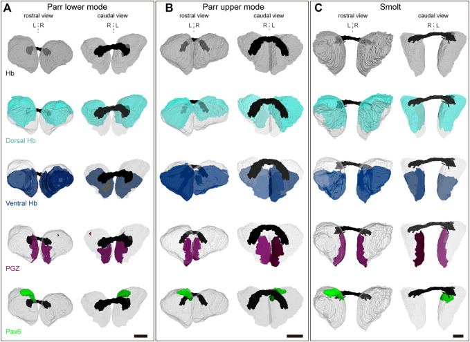
FIGURE 5 3D reconstructions of habenulae. (A–C) show 3D reconstructions of habenulae respectively in a parr lower mode, parr upper mode and smolt, with rostral and caudal views on the left and on the right as indicated. The whole habenulae are shown in grey (first line), the dorsal habenulae (including the pax6 dorsal left nucleus) in light blue (second line), the ventral habenulae (comprising kiss1 and sox1b positive territories) in dark blue (third line), the posterior growth zone (PGZ) in purple (fourth line) and the pax6 left dorsal nucleus in green (fifth line). The habenular commissure is in black. A dotted vertical line indicates the midline. Abbreviations: Hb, habenulae; L, left; R, right. Scale bars = 150 μm.