
|
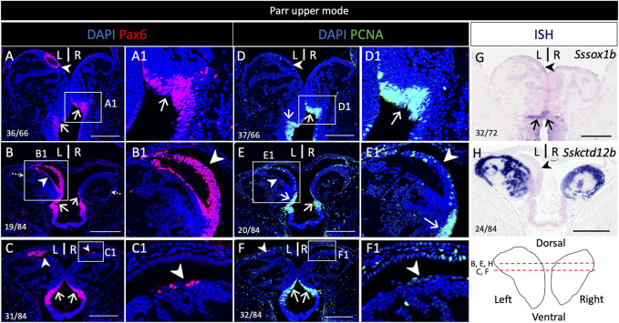
FIGURE 4 Pax6 expression in neural progenitors and a dorsal neuronal territory in the habenulae of Atlantic salmon. (A–H) show sections of a parr upper mode following IHC using antibodies directed against pax6 (A–C), PCNA (D–F) and following ISH using probes for Sssox1b (G) and Sskctd12b (H). (A,D,G) are transverse sections at the level indicated by dotted arrows in (B), (B–C,E,F,H) are horizontal sections at the level indicated by red lines on the front view of habenulae schematized in the bottom right panel. (A1–F1) are higher magnification views of the territories boxed in (A–F). Arrowheads point towards the left-restricted dorsal nucleus and a smaller, right-sided, dorsal population of clustered cells positive for pax6 but negative for PCNA. Thin arrows point towards posterior and ventral neural progenitors. Same abbreviations as in Figure 1. Scale bars = 200 μm.