
Fig. 6
- ID
- ZDB-IMAGE-221210-6
- Publication
- Walker et al., 2021 - Agrin/Lrp4 signal constrains MuSK-dependent neuromuscular synapse development in appendicular muscle
- All Figures
- Figures for Walker et al., 2021
|
Fig. 6
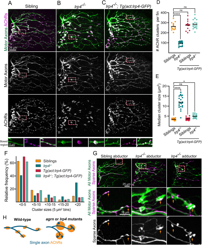
Lrp4 is required for correct axon innervation and AChR patterning in the pectoral fin. (A-C) Abductor muscle innervation in the pectoral fin in 120 hpf larvae expressing Tg(mnx1:GFP) to label motoneurons and stained with α-Btx to label AChRs. (A) lrp4 siblings exhibit innervation patterns with numerous small AChR clusters. (B) lrp4 mutants exhibit abnormal swellings in an innervation pattern directly opposed to that of large AChR clusters. (C) Expression of lrp4-GFP in muscles of lrp4 mutants is sufficient to rescue the mutant innervation pattern. Boxes outline the regions shown in insets. (D,E) Quantification of the number of AChR clusters per fin (D) and the median cluster size per fin (E). (F) Histogram of the distribution of AChR cluster sizes across all animals quantified (5 μm2 bins). n=15 (siblings), 23 (lrp4 mutants), 16 [siblings plus Tg(act:lrp4-GFP)], 20 [lrp4 mutants plus Tg(act:lrp4-GFP)]. (G) Sparse labeling of axons injected with mnx1:mKate, with all motor axons labeled with Tg(mnx1:GFP). Sparse labeling does not label entire ‘swelling’ but axon ends appear bulbous, indicating that multiple axons contribute to the abnormal innervation swellings in lrp4 mutants. Orange arrows indicate axon endings. Boxed indicate the regions magnified below. n=11/12 (sibling fins), 7/7 (mutant fins). (H) Schematic summarizing axonal organization and AChR clusters. ns, not significant. *P<0.05, ****P<0.0001, one-way ANOVA with Dunnett's multiple comparisons test.