
IMAGE
Fig. 4
- ID
- ZDB-IMAGE-221210-4
- Publication
- Walker et al., 2021 - Agrin/Lrp4 signal constrains MuSK-dependent neuromuscular synapse development in appendicular muscle
- All Figures
- Figures for Walker et al., 2021
Image
|
Figure Caption
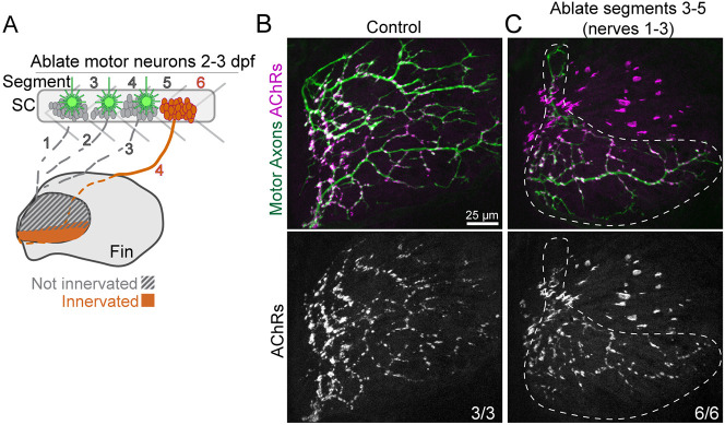
Fig. 4
Pectoral fin muscles are predisposed to form large AChR clusters. (A) Motoneuron cell bodies from spinal cord (SC) segments 3-5 were laser ablated at 2 and 3 dpf to prevent motor axon innervation of the dorsal pectoral fin. (B) Pectoral fins from control Tg(mnx1:GFP) larvae stained with α-Btx to label AChRs. (C) Pectoral fins from Tg(mnx1:GFP) WT larvae after motoneuron ablation at 120 hpf (5 dpf). The ventral fin is innervated (white-dashed region) with input from unablated nerve 4 (segment 6). The non-innervated dorsal region of the fin has enlarged AChR clusters.
Acknowledgments
This image is the copyrighted work of the attributed author or publisher, and
ZFIN has permission only to display this image to its users.
Additional permissions should be obtained from the applicable author or publisher of the image.
Full text @ Development