
Fig. 2
- ID
- ZDB-IMAGE-221210-2
- Publication
- Walker et al., 2021 - Agrin/Lrp4 signal constrains MuSK-dependent neuromuscular synapse development in appendicular muscle
- All Figures
- Figures for Walker et al., 2021
|
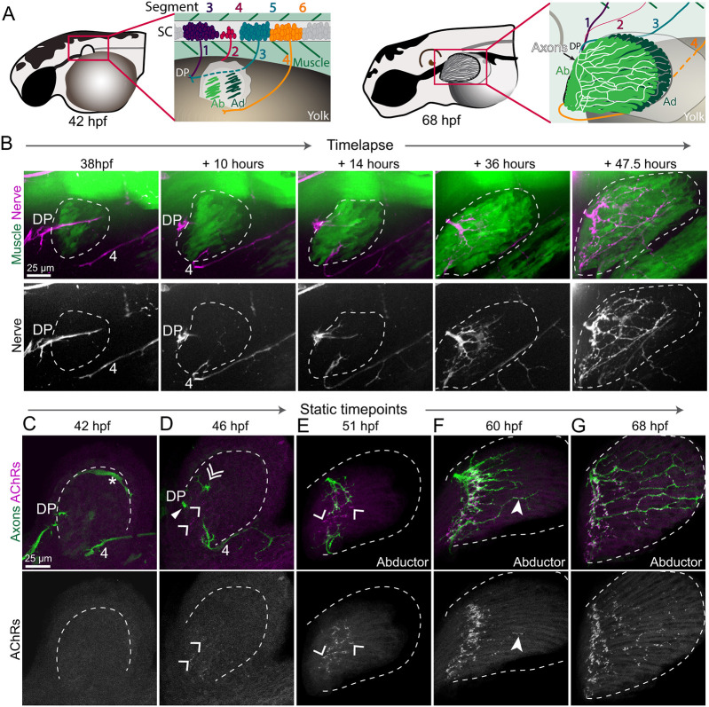
Fig. 2
Development of pectoral fin innervation. (A) Schematic of zebrafish larvae at 42 hpf (high-pec stage) and 68 hpf (pec fin stage). The boxed regions indicate the areas highlighted in the insets, showing motoneurons from spinal cord (SC) segments 3-6 and their corresponding nerves (1-4) projecting to the dorsal plexus (DP) to innervate the abductor (Ab) and adductor (Ad) muscles of the pectoral fin. (B) Maximum projection stills from time-lapse imaging of Tg(α-actin:GFP);Tg(Xla.Tubb:DsRed) larvae showing muscles and axons. Dashed lines outline pectoral fin musculature. Axons converge at the DP prior to innervating nascent muscle fibers. As muscle fibers elongate, the axonal innervation pattern elaborates (n=7 WT fins). Static time points of Tg(mnx1:GFP) larvae stained with α-Btx to label AChRs. Nerve 4 is indicated by ‘4’. (C) At 42 hpf, the pectoral fin bud is still lateral to the DP; thus, axons and muscles are not yet in the same plane. Asterisk indicates vasculature also labeled by mnx1:GFP. (D) Axons have just grown past the DP. Ab axons are indicated by the filled triangle, Ad axons are indicated by the double arrowhead and aneural AChR clusters are indicated by single arrowheads. (E-G) Ab innervation only. Axons occupy prepatterned clusters and induce new AChR clusters as they grow throughout the fin. Filled arrowhead indicates a fin axon branch already associated with AChR clusters. Single arrowheads indicate aneural AChR clusters. n=5-10 pectoral fins per time point for C-G.