
|
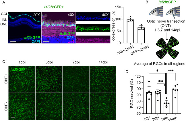
Fig. 1
Zebrafish RGCs are preserved after optic nerve transection. (A) Immunolabeling of RGCs in the GCL with zn-8 (magenta) in the adult isl2b:GFP (green) retinae. Shown are a retinal cross section (20×) and high magnification view of the GCL (40×). Quantification revealed that ∼65% of DAPI (blue)-stained RGCs were isl2b:GFP+ (n=xxx). Scale bars: 100 μm (20×); 50 μm (40×). (B) Overview of ONT and RGC survival analyses. (C) Images of flat-mount retinae at 1, 3, 7 and 14 dpi. Scale bar: 50 μm. (D) RGC survival percentages at 1, 3, 7 and 14 dpi (n=6/day). Graphs show mean±s.d.; *P<0.05; **P<0.01; ***P<0.001; Kruskal–Wallis ANOVA with Dunn's multiple comparisons. Data derived from three experiments.